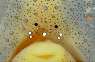
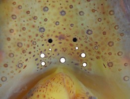
Table. Ventral view of the funnel-groove region of A. felis. Top tier - Funnel groove showing photophores of funnel groove and surrounding tissue. Middle tier - In the photograph, white dots placed over the "white" photophores of the funnel groove. Black dots placed over the most posteromedial "red" photophores of the ventral head to mark the mark the anterolateral edges of the funnel groove. Bottom tier - Same pattern of dots as in photographs. Colors, lines - Aid in comparison of patterns between species. Images by R. Young.
| Mature female, 36 mm ML | Mature female, 37 mm ML |
|---|---|
 |  |
 |  |
 |  |


 Go to quick links
Go to quick search
Go to navigation for this section of the ToL site
Go to detailed links for the ToL site